
Analysis, comparison, visualization of contact residues & interfacial waters of antibody-antigen structures/models
|
 |
|
|
|
|
|
|
|
|
|
|
|
|
|
|
|
|
|
Antibodies are currently the fastest growing class of therapeutics (Fischman and Ofran, Curr. Opin. Struct. Biol., 2018). For example, the antibody drugs Humira, Opdivo, Keytruda, Herceptin, Rituxan, and Avastin that our AppA web server performed structural analysis in the below figure are currently in the top 10 pharmaceutical products on the market in 2018. The study of contact residues and interfacial waters of antibody-antigen structures could help in understanding of the principles of antibody-antigen interactions as well as provide excellent means to improve affinity. Given the rapid pace with which new antibody-antigen structures are deposited in the PDB (4095 protein antibody–antigen structures as of 28 Mar 2022), it is crucial to have computational tools to analyze contact residues and interfacial waters, and investigate them for similarities at different levels. In this study, we have developed AppA a web server that is capable for analysis of contact residues and interfacial waters of antibody-antigen complexes as well as comparison of their 3D structures. AppA identifies and analyzes contact residues and interfacial waters of antibody-antigen structures/models by using the computational methods in our previous study (Nguyen et al., Bioinformatics, 2017). Our study has demonstrated the important roles of contact residues and interfacial waters to mediate the interactions of antibodies and antigens (Nguyen et al., Bioinformatics, 2017). AppA compares the 3D structures of contact residues by optimizing the CLICK algorithm (Nguyen and Madhusudhan, Nucleic Acids Res., 2011). CLICK has been extensively benchmarked to other popular methods for protein structural alignments and comparison of binding sites (Nguyen and Madhusudhan, Nucleic Acids Res., 2011, Brown et al., Bioinformatics, 2015, and Garma et al., Proteins, 2016). Various examples showcase the utility of AppA for analysis of structures of contact residues and interfacial waters that could help understanding of how antibody interaction with antigen and suggest mutation of contact residues to improve affinity. In addition, AppA will automatically identify and calculate hydrogen bonds, van der Waals, hydrophobic, ionic interactions in the antibody-antigen interface, and interfacial waters of new antibody-antigen structures deposited in the PDB every month. The list of antibody-antigen complexes is also updated every month from the PDB. |
|
|
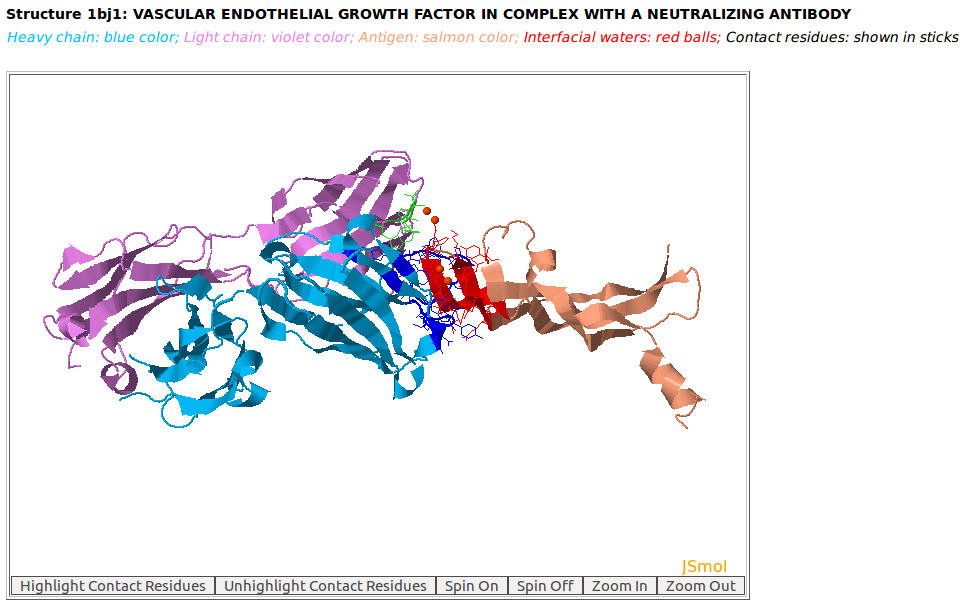
AppA displays the 3D rendition of the antibody drug Avastin (generic name: bevacizumab, PDB code: 1BJ1) with heavy chain (H, blue color), light chain: (L, violet) and the antigen VEGF (Human Vascular Endothelial Growth Factor; chain W, salmon color) and interfacial waters (red balls) using JSMol (https://jmol.sourceforge.net/). The contact residues of Avastin and VEGF are highlighted in sticks. |