Comparison of RNA 3D Structures

Introduction

Examples

Benchmark

Run Rclick

References

Contacts

Help

Structural alignment of two RNA aptamer structures 1OOA:D and 2JWV:A by Rclick

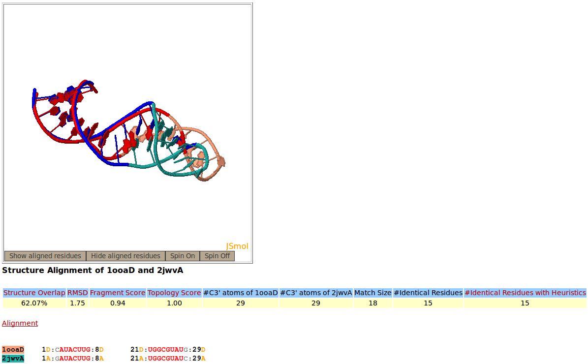
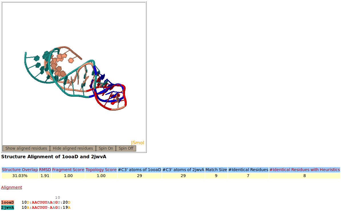
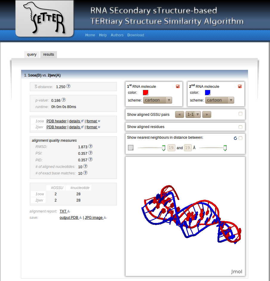
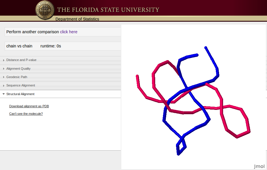
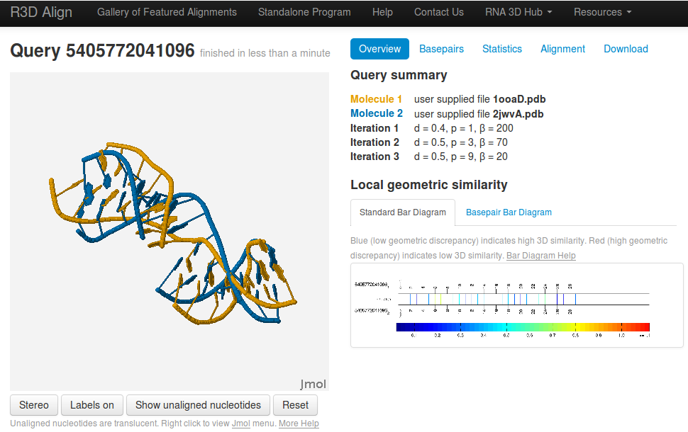
Snapshot of the Rclick showing the first alignment of two RNA aptamer structures, PDB codes 1OOA:D (salmon), and 2JWV:A (green). The superimposed residues of 1OOA:D and 2JWV:A are shown in red and blue,respectively. The regions, spanning residues 1-8 and 21-29 are aligned with one another. Because of a conformational change, the regions of residues 10-20 do not align structurally.

With conformational change, Rclick shows the second alignment of the regions of residues 10-20 of 1OOA:D and 2JWV:A


[back to top]


Structural alignment of two RNA aptamer structures 1OOA:D and 2JWV:A by SARA


[back to top]


Structural alignment of two RNA aptamer structures 1OOA:D and 2JWV:A by ARTS


[back to top]


Structural alignment of two RNA aptamer structures 1OOA:D and 2JWV:A by SETTER


[back to top]


Structural alignment of two RNA aptamer structures 1OOA:D and 2JWV:A by RASS


[back to top]


Structural alignment of two RNA aptamer structures 1OOA:D and 2JWV:A by R3DAlign


[back to top]